Table of Contents
- 1. Introduction: The AI Era in Radiology
- 2. What Is Medical Imaging and Why Does It Matter?
- 3. How Does AI Work in Medical Imaging?
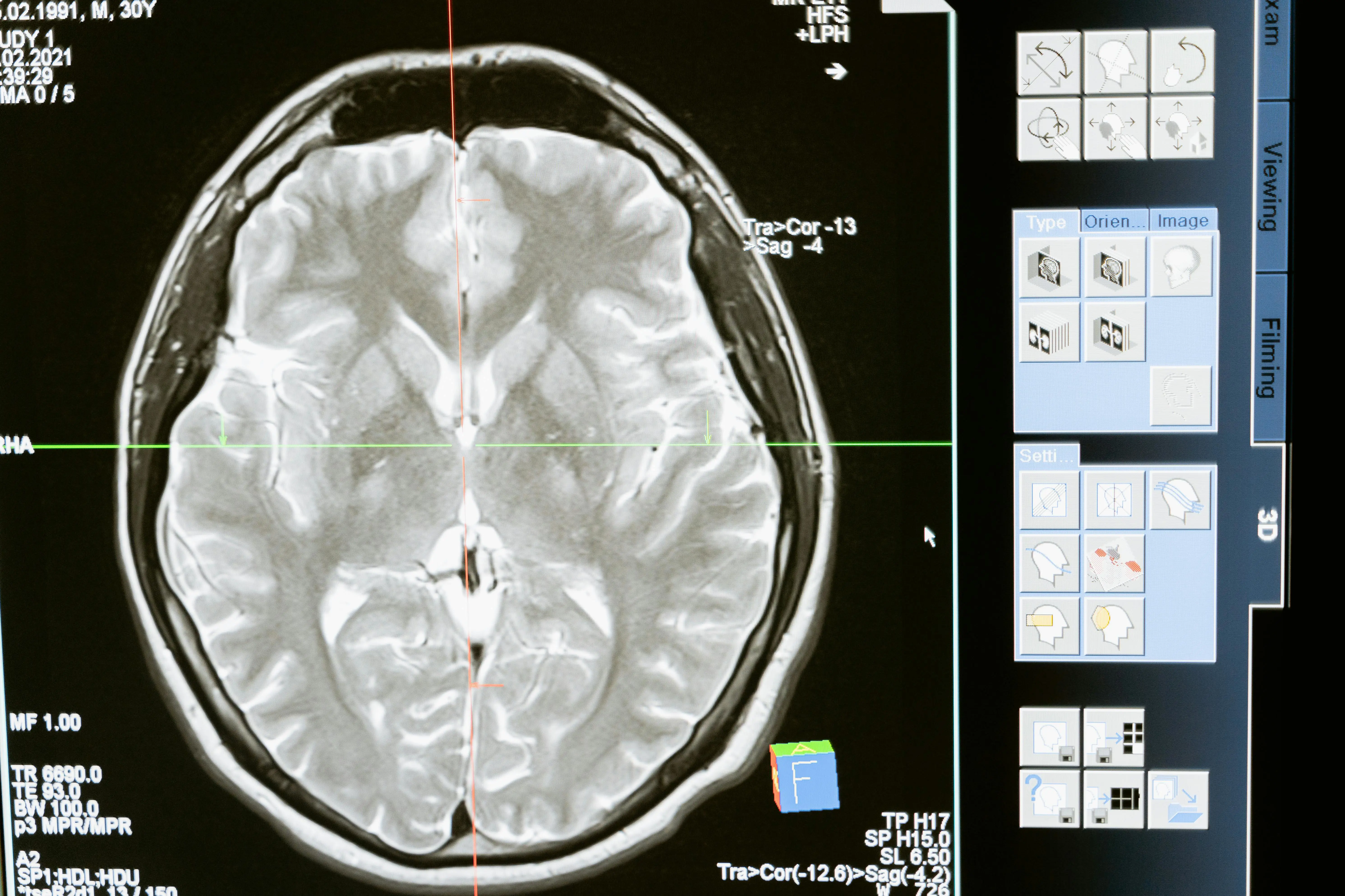
- 4. AI in CT, MRI, and X-Ray Analysis
- 5. Pathology Detection and Early Diagnosis
- 6. AI in Early Cancer Screening
- 7. FDA-Approved AI Tools
- 8. Accuracy Rates and Clinical Evidence
- 9. Integration into Radiologist Workflow
- 10. Future Perspective
- 11. Frequently Asked Questions (FAQ)
1. Introduction: The AI Era in Radiology
Medical imaging stands as one of the fundamental pillars of modern healthcare. Billions of medical images are generated worldwide each year, and this number continues to grow exponentially. Radiologists invest tremendous effort in detecting diseases, making diagnoses, and planning treatments within this enormous volume of data. This is precisely where artificial intelligence technologies emerge as a transformative force in the field of radiology.
As of 2026, the AI-powered medical imaging market has surpassed $7 billion in value. The FDA has granted clearance to over 800 AI-based medical devices and software, with the vast majority concentrated in the radiology domain. These figures clearly demonstrate the enormous impact AI is having on the healthcare sector.
Key Insight
Artificial intelligence is designed not to replace radiologists but to accelerate their diagnostic processes and increase accuracy rates. AI can be thought of as the radiologist's "super-powered eyes."
In this comprehensive guide, we will explore AI applications in medical imaging in detail, examining its role in CT/MRI/X-ray analysis, its success in early cancer screening, FDA-approved tools, and the future of radiology.
2. What Is Medical Imaging and Why Does It Matter?
Medical imaging refers to the use of various technologies to visualize the internal structures of the human body. These technologies play an indispensable role in disease diagnosis, treatment planning, and follow-up monitoring.
Core Imaging Modalities
Approximately 3.6 billion medical imaging procedures are performed globally every day. This immense volume creates a workload that pushes the limits of human visual perception, making AI support increasingly important with each passing day. When you consider that radiologists evaluate an average of 50 to 100 cases per day, AI's contribution to this process becomes much clearer.
3. How Does AI Work in Medical Imaging?
The success of artificial intelligence in medical imaging is largely based on deep learning algorithms. Convolutional Neural Networks (CNNs) in particular demonstrate extraordinary performance in image recognition and analysis tasks.
Deep Learning Architecture
AI models used in medical imaging are typically trained through the following stages:
- Data collection: Hundreds of thousands of labeled medical images are compiled. These images are annotated by expert radiologists.
- Preprocessing: Images are normalized, resized, and data augmentation techniques are applied.
- Model training: The model is trained using architectures such as CNN, U-Net, ResNet, or Vision Transformers.
- Validation: Model performance is tested with datasets outside the training set.
- Clinical validation: Success rates are measured through prospective studies in real clinical settings.
Core AI Techniques Used
Segmentation: Determines pixel-level boundaries of organs, tumors, or lesions. The U-Net architecture is considered the gold standard in this area. This enables radiologists to evaluate suspicious regions with millimeter precision.
Classification: Categorizes findings in images as benign or malignant. In this process, pre-trained models are fine-tuned with medical data using transfer learning methods.
Object detection: Automatically locates and marks findings such as anomalies, nodules, or calcifications in images. Architectures like YOLO and Faster R-CNN are widely used for this task.
Important Warning
AI models are not perfect and should always be used under the supervision of expert radiologists. Artificial intelligence should be positioned as a decision support system; the final diagnosis should always remain the clinician's responsibility.
4. AI in CT, MRI, and X-Ray Analysis
AI in CT (Computed Tomography) Analysis
CT scans provide cross-sectional images of the body, enabling detailed examination of internal organs. Artificial intelligence is revolutionizing CT imaging in the following areas:
- Lung nodule detection: AI algorithms can detect nodules as small as 3mm with over 95% accuracy. This capability is critically important for catching early-stage lung cancer.
- Coronary calcium scoring: Automatically measures calcifications in coronary arteries for cardiovascular risk assessment.
- Stroke detection: Detects large vessel occlusion (LVO) in brain CT images within seconds, reducing time to emergency intervention. LVO algorithms play a life-saving role in this area.
- Liver lesions: Performs automatic segmentation and characterization of liver tumors such as hepatocellular carcinoma.
AI in MRI (Magnetic Resonance Imaging) Analysis
MRI offers superior performance in soft tissue contrast. AI's contributions to the MRI field include:
- Brain tumor segmentation: Volume measurement and growth tracking of glioblastoma and other brain tumors have been automated with AI.
- Prostate cancer detection: AI models compatible with PI-RADS classification can identify suspicious lesions with over 90% sensitivity.
- Cardiac MRI analysis: Left ventricular ejection fraction, myocardial wall thickness, and perfusion analysis are automatically calculated.
- Accelerated MRI: AI enables high-quality image reconstruction from undersampled k-space data, reducing scan time by up to 75%.
AI in X-Ray Analysis
X-ray is the most widely used and accessible imaging method. AI is making a significant difference in X-ray analysis in the following areas:
- Chest X-ray analysis: Simultaneously screens for more than 14 pathologies including pneumonia, tuberculosis, pleural effusion, cardiomegaly, and pneumothorax.
- Fracture detection: Detects hand, wrist, and hip fractures with accuracy rates reaching 97%. Particularly advantageous for catching occult fractures.
- Bone age assessment: Automatically performs bone age determination in pediatric patients.
- Dental X-ray: AI-assisted analysis for cavity detection, periapical lesions, and periodontitis is becoming increasingly widespread.
5. Pathology Detection and Early Diagnosis
One of the most critical contributions of AI in medical imaging is enabling early and accurate detection of pathologies. The capacity to capture subtle changes that the human eye might miss makes AI an indispensable assistant in the diagnostic process.
Automated Anomaly Detection
AI systems are trained on millions of normal and abnormal images to detect deviations from normal anatomy. This gives them the ability to prioritize cases that most require the radiologist's attention. For example, in an emergency department setting, the system can flag life-threatening findings such as intracranial hemorrhage or pneumothorax within seconds and move them to the top of the worklist.
Deep learning models demonstrate high performance in detecting the following pathologies in particular:
Triage and Prioritization
AI-powered triage systems significantly improve the efficiency of radiology workflows. Images containing urgent findings are automatically moved to the top of the worklist, while cases with normal findings are queued for later evaluation. This approach reduces diagnosis time in critical cases by an average of 60-70%.
6. AI in Early Cancer Screening
Cancer is one of the most significant health challenges worldwide, and early detection dramatically improves survival rates. AI is delivering revolutionary improvements in cancer screening programs.
Breast Cancer Screening
The use of AI in mammography screening has significantly increased breast cancer detection rates. Studies show that AI-assisted screening reduces false negative rates by 30-40% compared to single-radiologist evaluation. Furthermore, AI's performance is particularly notable in women with dense breast tissue, as dense tissue can cause lesions to be missed with conventional methods.
Lung Cancer Screening
Low-dose CT lung cancer screening is a life-saving approach for high-risk individuals. AI algorithms not only detect lung nodules but also assess the malignancy risk of nodules, reducing unnecessary invasive procedures. Google Health's AI model achieved 5% higher accuracy than a panel of six radiologists.
Colorectal Cancer Screening
AI-assisted colonoscopy and CT colonography are becoming increasingly prevalent for detecting polyps and adenomas. Real-time AI assistants instantly flag small polyps that the endoscopist might miss, increasing the adenoma detection rate (ADR) by 14-30%.
Pro Tip
Early-stage cancer detection can raise the 5-year survival rate above 90%. AI-powered screening programs significantly increase this early detection potential. Participation in regular checkups and screening programs can save lives.
7. FDA-Approved AI Tools
The U.S. Food and Drug Administration (FDA) evaluates medical AI software under the SaMD (Software as a Medical Device) framework. As of 2026, the number of FDA-cleared radiology AI tools has exceeded 500. Here are some of the most prominent FDA-approved AI tools:
The common feature of these tools is that they are designed to integrate seamlessly into clinical workflows. Through direct integration with PACS (Picture Archiving and Communication System), radiologists can benefit from AI support without disrupting their existing work patterns.
In Europe, the approval process is conducted under CE marking. The regulatory landscape continues to evolve globally, with each region adapting its frameworks to accommodate the rapid development of AI-based medical software while maintaining patient safety standards.
8. Accuracy Rates and Clinical Evidence
AI's accuracy rates in medical imaging have been extensively researched in scientific literature. Numerous studies demonstrate that AI performs equivalently to or better than expert radiologists in specific tasks.
Comparative Performance Data
A comprehensive meta-analysis published in Nature Medicine examined the diagnostic accuracy of deep learning algorithms. Results showed that the average AUC (Area Under the Curve) of AI models was 0.94, close to the expert radiologist average of 0.91.
However, an important point to note is that the AI + radiologist combination consistently outperforms either AI alone or radiologist alone. This "human-machine partnership" approach forms the foundation of future radiology practice.
Clinical Study Results
- OPTIMAM study: AI-assisted mammography screening achieved a 17% reduction in false positive rates.
- CheXpert study: In chest X-ray analysis, AI achieved higher AUC values than 3 radiologists across 5 pathology categories.
- RSNA 2025 data: AI triage systems reduced average emergency radiology reporting time from 45 minutes to 12 minutes.
- Sweden MASAI trial: AI-assisted mammography screening increased cancer detection rates by 20% while reducing radiologist workload by 44%.
Key Insight
AI model performance is directly related to the quality and diversity of training datasets. Models trained on diverse datasets encompassing different ethnic groups, age ranges, and clinical scenarios produce more generalizable and equitable results.
9. Integration into Radiologist Workflow
Integrating AI into radiology practice is not merely a technological matter but also an organizational and cultural transformation process. Successful integration requires the evaluation of multiple factors in tandem.
PACS Integration
Modern AI solutions work in integration with hospital PACS systems via the DICOM protocol. Images are subjected to AI analysis the moment they are acquired, and results are automatically delivered to the radiologist's workstation. DICOM SR (Structured Report) and DICOM SC (Secondary Capture) formats are used to present findings in a standardized format.
Workflow Optimization
The radiologist workflow is optimized through AI integration in the following ways:
- Smart worklist management: Critical findings are automatically prioritized and notifications are sent to the radiologist.
- Pre-read support: AI marks suspicious areas to direct the radiologist's attention and reduce the risk of oversight.
- Automated measurements: Organ sizes, lesion diameters, and volume calculations are performed automatically, shortening reporting time.
- NLP-assisted reporting: AI automatically structures reports dictated by the radiologist and compares with prior studies.
- Follow-up recommendations: Standardized guideline-compliant follow-up recommendations are automatically generated based on detected findings.
Training and Adaptation
Comprehensive training programs are required for radiologists to effectively use AI tools. Understanding AI's strengths and weaknesses, correctly interpreting false positive and false negative results, and critically evaluating AI recommendations are fundamental components of successful integration. The inclusion of AI education in medical school curricula is also becoming increasingly common.
10. Future Perspective
The future of AI in medical imaging holds exciting developments. Technological advances, new application areas, and changing healthcare paradigms point to a fundamental transformation in radiology over the next decade.
Emerging Technologies
- Foundation models: The adaptation of general-purpose large language models (LLMs) to medical imaging will enable the development of models that deliver high performance even with limited data. Models like Google's Med-PaLM and Microsoft's BiomedCLIP are pioneering this field.
- Multimodal AI: Analyzing imaging data alongside clinical notes, laboratory results, and genomic data will offer more comprehensive and personalized diagnostic capabilities.
- Federated learning: Multiple institutions will be able to collaboratively train AI models while ensuring patient data never leaves the hospital. This approach balances data privacy with model performance.
- Explainable AI (XAI): AI models that visually explain the rationale behind their decisions will increase radiologists' trust and bring transparency to the clinical decision-making process.
The Evolving Role of Radiology
Artificial intelligence will not eliminate the role of radiologists but rather enrich it. Radiologists will be freed from routine and repetitive tasks, allowing them to focus on more complex cases, multidisciplinary decision-making processes, and patient communication. The concept of the "augmented radiologist" best describes the future of radiology practice.
Within the next 5 to 10 years, AI is expected to become an integral part of standard care practice in radiology. In developing countries, particularly in regions with radiologist shortages, AI will serve as a critical tool for democratizing access to healthcare services.
Conclusion
AI-powered medical imaging is experiencing one of the most exciting transformations in the healthcare sector. With early and accurate diagnosis, optimized workflows, and the vision of personalized medicine, AI will continue to shape the future of radiology.
11. Frequently Asked Questions (FAQ)
Will AI replace radiologists?
No, artificial intelligence is designed to enhance radiologists' capabilities, not replace them. AI assists radiologists as a decision support system. Tasks such as clinical context evaluation, patient communication, and multidisciplinary decision-making will continue to require human expertise. The future of radiology will be shaped by the difference between radiologists who use AI and those who do not.
How accurate is AI-powered medical imaging?
While AI model accuracy varies by application area, it generally falls within the 90-98% range. Sensitivity rates reach 96-99% for diabetic retinopathy detection, 94-97% for lung nodule detection, and 95-98% for intracranial hemorrhage detection. The highest accuracy is achieved when AI and radiologists work together.
How does AI protect patient privacy in medical imaging?
Patient privacy in AI systems is protected through various technical and legal measures. Data is anonymized to remove personal information, federated learning enables model training without data leaving the hospital, and regulations like HIPAA and GDPR restrict data use within legal frameworks. Additionally, end-to-end encryption and access controls ensure data security.
Is early cancer detection with AI really possible?
Yes, AI's success in early cancer detection has been proven through scientific studies. In areas such as breast cancer in mammography, lung cancer in low-dose CT, and skin cancer in dermoscopy, AI can detect cancer at early stages that the human eye might not catch. The MASAI study conducted in Sweden showed that AI-assisted screening increased cancer detection rates by 20%.
What is the difference between deep learning and traditional image processing?
Traditional image processing uses manually defined features (edge detection, thresholding, morphological operations), while deep learning uses hierarchical features automatically learned from data. Deep learning can capture much more complex patterns with millions of parameters and generally achieves significantly higher accuracy rates compared to traditional methods.
Is it mandatory to use FDA-approved AI tools?
AI software used for clinical decision-making requires FDA clearance in the U.S., CE marking in the EU, and equivalent regulatory approval in other regions. This requirement may not apply for research purposes; however, the use of approved tools is strongly recommended for patient safety.
How long does it take to integrate AI into a radiology department?
Integration timelines vary depending on the complexity of the AI solution, existing IT infrastructure, and institutional readiness. A basic AI triage tool can be deployed within 2-4 weeks, while a comprehensive AI suite with multiple applications may take 3-6 months for full integration, including testing, validation, and staff training phases.